

Entry #02
"Island in a Steel Sea"
Artist's Statement: In this piece, I was deeply drawn to the isolation of the brittle area around what looks like waves of ductile dimpling. The steely waves then made me think of water and the isolation of a deserted island. The coloring I utilized then made this abundantly clearer as if looking at a simple map of an island in a sea of steel. While Happy Valley may not have a sea or an ocean coast, there are many lakes in the area and some even have islands of their own! These unexplored territories and habitats that this image reminded me of further connected me, my home, and this piece in my mind and my heart. These colors particularly standout to me as well as the general shape and topography of the image and I hope that they make the piece pop to the audience while reminding them of unfound, isolated lands as well.

Entry #03
"Made from Scratch: Scratch Induced Birefringence in Conjugated Polymer Indacenodithiophene-co-benzothiadiazole (IDT-BT)"
Artist's Statement: This unedited image calls to mind the beautiful blue Morpho menelaus butterfly, which has strikingly blue wings that are delicate but powerful. Like the Morpho menelaus butterfly, this conjugated polymer thin film gets its color from its unique nanoscale structural properties and not because it is pigmented.

Entry #04
"Graphene x Andy Warhol"
Artist's Statement: I remember seeing the MVC winners from past years hanging in Steidle during my campus tour and thinking, "Wow, I hope one day I will be able to do something like that." Now here I am, 2 years later, submitting my own image for MVC 13. Andy Warhol is well known for his pop art, and since I grew up in Pittsburgh like him, I couldn't resist drawing the comparison to my image. My piece is authentic, straight out of the Nano Scope software. I know it will be hard to compete with the extraordinary computational and SEM images, but I firmly believe I have made the best possible submission I could with the tools and knowledge I have.

Entry #05
"Formation of a discotic liquid crystal: Nucleation, growth, and coalescence of mesophase spheres driven by the principles of chemistry and physics into a visual narrative of science with line, shape, color, texture, and space as shown in a polarized-light micrograph"
Artist's Statement: This is a microscope image of a semi-coke obtained from carbonization of a petroleum feedstock in a laboratory reactor. To the naked eye, it appears black, similar to coal. “The color black relates to the hidden, the secretive and the unknown, and as a result it creates an air of mystery.” A polarized-light microscope may uncover this mystery and liberate the colors blue, yellow, and magenta by the interaction of polarized-light with anisotropic carbons. Capturing all three stages of mesophase development in just one image offers a powerful visual narrative of the dynamic process with changes in shape, color, and texture of objects from the birth of mesophase spheres (nucleation) on the right of the image to their growth and interaction with others (coalescence), and into their full maturation into anisotropic domains on the far left. In addition, the memory of these interactions is retained with patches of yellows and blues remaining from individual spheres that were coalesced.

Entry #06
"Rigidity of the extracellular matrix surrounding the cell promotes Epithelial-Mesenchymal Transition"
Artist's Statement: My research is based on the big-picture of understanding how the mechanical properties of a tissue regulate cell signaling pathways. If we can better understand how cells behave in response to tissue mechanics, we can develop strategies to target the signaling pathways and in turn, negate pathological conditions such as cancer and fibrosis. My current work involves studying the effect of modulus of the cell’s extracellular matrix on chromatin architecture and how it affects gene transcription machinery during the process of Epithelial-Mesenchymal Transition (EMT). The picture I am trying to convey with the help of my image submission is that mechanical stimuli can independently regulate the expression of proteins in epithelial cells during the process of EMT. Hence, I combined fluorescence images taken for EMT-induced epithelial cells that were seeded on increasing matrix rigidities to show the audience how the increase in modulus enhances EMT-induced protein expression in cells

Entry #07
"If Plants Grew like Lithium Niobate Crystals, then I’d Plant a Garden of Sunflowers"
Artist's Statement: While black and white electron images are quite possibly the farthest thing from a colorful garden, the radially growing lithium niobate crystals wedged between potassium niobium silicate oxide reminded me of blossoming flowers. As a dabbling gardener myself, I usually focus my energy on growing vegetables. But no matter how hard I try, stray sunflowers always seems to pop-up in the veggie garden. Always appearing with nothing more than soil and sun, sunflowers represent the all-to-easily formed lithium niobate crystals in my SiO2-Nb2O5-Li2O-K2O glass system. Just like sunflowers grow in the garden, lithium niobate crystals grow in my glass-ceramic, producing a beautiful bouquet of flowering crystals in a garden of glass.

Entry #09
"Heavenly Patterns: Widmanstätten Microstructure in Additively Manufactured Duplex Stainless Steel"
Artist's Statement: I still remember the day I first used a microscope to look at onion cells back in middle school. I was astonished at what lies beyond the view of the naked eye. Fortunately, till this day I am still in awe of the world found under a microscope. Microscopy helps us understand much more about material systems and how new sustainable means of manufacturing influence them. Additive manufacturing is a gateway to new discoveries that will eventually improve upon traditional manufacturing methods resulting in more cost-effective and advanced materials with better properties. As the perseverance rover recently landed on the surface of Mars, one day the next mission will be carrying a 3D printer capable of printing materials on the spot aiding the journey of interplanetary travel.

Entry #10
"Interconnected microporous structure of tung oil and poly(ε-caprolactone) semi-interpenetrating polymer networks"
Artist's Statement: This picture observed by scanning electron microscopy. I was very surprised to see a wonderful black and white microscale morphology of semi-interpenetrating polymer networks that you may be able to see similar large-scale pictures when you are diving in the deep ocean.

Entry #12
"Never-ending Friendship: Titanium(IV) isopropoxide (TTIP) decomposition on SrO terminated SrTiO3"
Artist's Statement: As a child of immigrants who moved from the former Yugoslavia to Turkey and now an international student in the US, the concept of friendship has been very meaningful for me my entire life. Wherever I have been, the friends who received me with open arms broadened my horizons and motivated me to discover more outside my little family. Likewise, there is a never-ending friendship between TTIP and SrO-terminated surface where TTIP discovers itself as well as its environment. That way, this tiny TTIP molecule is finally able to be a harmonious part of the atomic community in which it is more than welcome.

Entry #13
"Microscopic Magnesium Meteor"
Artist's Statement: Beautifully falling into the unknown. This crystal appears to fly across a world that is strange from itself. Like a magnesium-rich meteor falling to Earth, it lights up and lets the world know of its arrival. I was inspired to represent this crystal as a meteor because the odds of this crystal forming are comparable to a meteoroid entering Earth’s atmosphere. Space is infinitely large and the likelihood of any specific meteoroid reaching Earth is improbable, just like the probability of this lone magnesium crystal forming in its boron-rich environment.

Entry #14
"Gold droplets on graphene leaf"
Artist's Statement: My passion for photography always makes me to make an analogy between my work to the daily life. This work explores the relationship between scientific output of my experimental work of gold metal intercalation on graphene surface to the the real-life water droplets on leaves. As the real scientific output image is a colorless, I wanted them to turn around into a colored daily life image that my audience is familiar with. For that I have retouched them using photoshop software and made a colorful one.

Entry #17
"Mixed Chromium Oxide in the Shape of a Skull"
Artist's Statement: As many discoveries are sometimes made, I came upon this surface defect entirely by chance during the characterization of this sample with Raman spectroscopy. Imagine my surprise while collecting a spectrum to see a skull looking back at me! The morphology of our samples created through laser-induced thermal synthesis have been well studied through other methods such as scanning electron microscopy and profilometry. This image reminds us to always keep our eyes peeled for discovery in our research, whether it’s noticing a unique surface feature while collecting a spectrum or finding new properties in a material.

Entry #19
"Multimetal Particles Sintered to Glass Substrate"
Artist's Statement: When I first saw this film, I was vaguely reminded of an abstract night sky with the gray substrate resembling the vast space that was then dotted with stars (Fe/Co/Ni oxide particles in reality). As I continue my research in the Zarzar group at Penn State, I am thankful of these moments where I can repeatedly experience the awe and wonder of scientific observation.

Entry #20
"Dendrites in a Battery"
Artist's Statement: Light like a feather and delicate like a fern? Do not let the looks fool you. These metal dendrites grown from battery cycling will penetrate the separator, short-circuit the battery, and cause the battery to overheat and, in some instances, catch on fire. This image inspires me because of the soft, delicate appearance in contrast to the material's stiff nature, a candy to eyes but a poison to batteries.

Entry #21
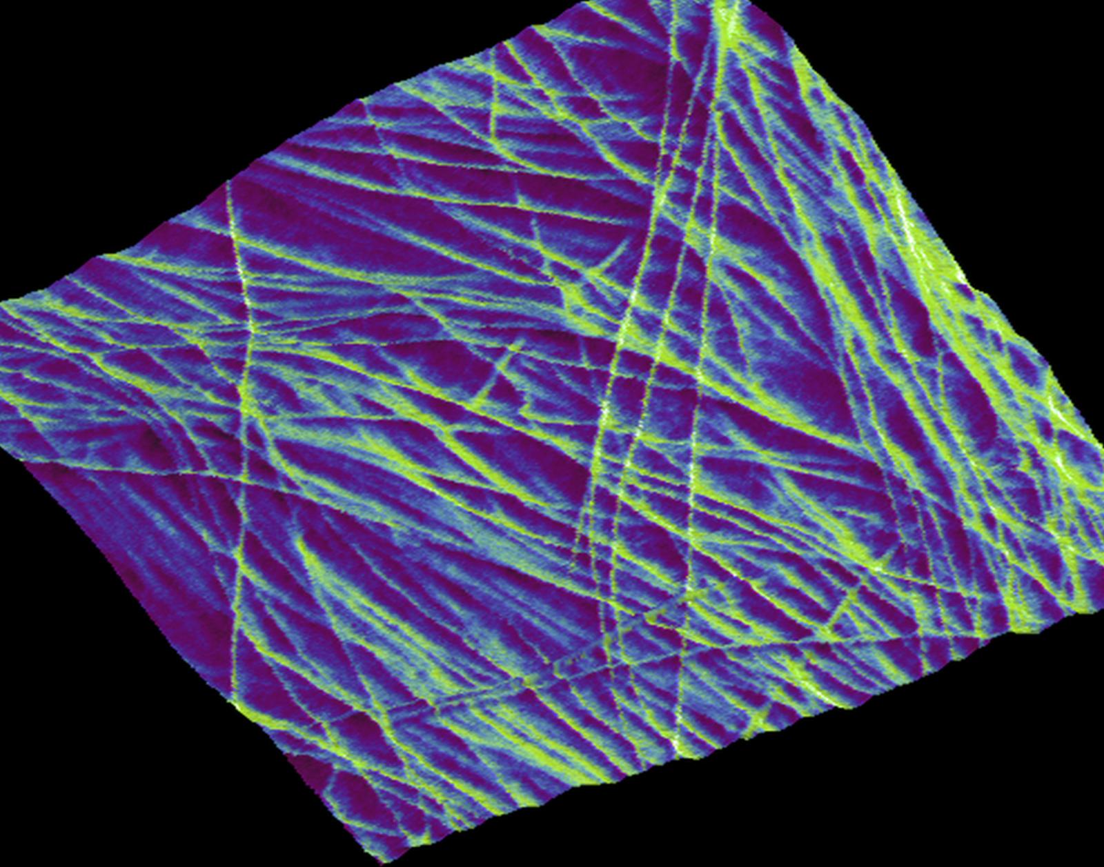
"Newly deposited cellulose of primary cell wall"
Artist's Statment: The primary cell walls in higher plants are an intricate network of cellulose, pectins, and other polysaccharides that provide a rigid support to stand upright, yet it becomes flexible when the plant is growing. The motivation behind this image is to capture how the cellulose is orientated in the wall using a non-destructive, native-condition imaging technique known as Atomic Force Microscopy (AFM). This imaging technique allows one to capture features as small as 2 nanometers in diameter, well below the resolution of confocal microscopy. My goals with this technique are to understand how the different components of the wall are formed and how they interact with each other. This image shows how intertwined the layers of cellulose are in the wall and how these patterns can allow for ‘movement’ of the wall to allow growth. I hope that when viewers see this image, they are curious about the plants around them, and want to learn more about how they are built and how they grow.
Entry #22
"Strength in Numbers: Cellulose Bundling in Plant Secondary Cell Walls Reinforces Individual Cells"
Artist's Statement: I chose this novel snapshot of cell wall formation because it depicts the bundling of cellulose in the plant secondary cell wall in a very palpable way. The formation of secondary cell walls is what allows plants to grow vertically. In this atomic force microscopy scan, the secondary cell wall bundles are evocative of tree trunks that grow upwards and then branch outwards. This web of cellulose bundles provides strength on the microscale in a way that mirrors the structure that they ultimately support on the macroscale.

Entry #23
"Different facets of unearthed carbides"
Artist's Statement: Carbides are hiding away from the melting pit and plotting a scheme to escape.

Entry #24
"Converting cost-effective polyacrylonitrile/poly(p-phenylene-2,6-benzobisoxazole) blend precursors to carbon fibers"
Artist's Statement: The traditional approach of producing polyacrylonitrile (PAN)-derived carbon fibers (CFs) is expensive, partially due to the rigorous control over the sequence of thermal treatments such as oxidative stabilization, carbonization, and graphitization. To this end, we propose the PAN/poly(p-phenylene-2,6-benzobisoxazole) (PBO) blend as a cost-effective CF precursor. And through our ReaxFF molecular dynamics (MD) simulations, we identify that PAN/PBO blends could be a promising alternative for PAN-based precursors, for they can decrease the total cost of CF production by eliminating oxidation process, having a relatively fast conversion rate, and having considerable all-carbon ring formation, comparable to that of oxidized PAN precursor. Based on this finding, a VMD image is made as to describing the process of converting a PAN/PBO blend precursor to clusters of all-carbon ring structures, followed by graphitic 3D or graphene-like 2D structures as the temperature is sufficiently high.

Entry #25
"Manifold of self-assembled nanostructures for a model copolymer obtained by unsupervised machine learning"
Artist's Statement: The data and rendering were created using all open-source tools: simulations in HOOMD-blue, data analysis in Python, and rendering in Blender. The colors and arrangements of particles were all computed automatically through molecular dynamics and the UMAP algorithm. The colors of particles communicate the local structure in the material while arrangements of simulation snapshots communicate global trends in the structural phase space. The act of generating this image therefore provides scientific insight on its own, by conveying (nearly) all possible nanostructures that can be self-assembled by this model copolymer under certain constraints. Rather than seeking a logical arrangement or interpretation of all these many simulations, automated workflows provide quantitative understanding of relationships between data. In turn, these workflows allow me to tackle extremely large design spaces and volumes of simulation data by reducing the data down to only a few key parameters.

Entry #26
"Electrochemical charging near a pseudocapacitive electrode surface"
Artist's Statement: With the growing needs for cleaner energy storage and conversion, realistic models of energy storage materials are needed to aid in the design of high-performance energy storage devices. Specifically, there is a need for high-power devices for use in electric/fuel cell vehicles. First-principles calculations with continuum solvent models allow us to model these materials systems in realistic conditions. The system pictured is a Ti3C2O2 pseudocapacitive electrode, suitable for high-power transportation applications. As the electrode charges, the response of the ions in the solvent, shown as colored contours above and below the electrode surface, is related to the capacitance of the electrode.

Entry #27
"Scanning electron micrograph of an etiolated Arabidopsis seedling"
Artist's Statement: A scanning electron micrograph of an etiolated Arabidopsis seedling (grayscale) growing in the darkness, just after it has shed its seed coat (orange). This electron micrograph is superimposed on a photograph of protein conjugated nano-gold labeled on peeled Arabidopsis seedings epidermal cell wall. This is the moment right before it would break free from the earth and reach toward the sunshine.

Entry #28
"The Silver Albacore"
Artist's Statement: A fun part of any cloudy day is always looking in the sky and trying to find figures in the clouds with friends. I compare the nanocubes I synthesize to looking at cloud formations; If you look long enough, you can see something fun in every single sample. The image was created with silver nanocubes, capped with polyvinylpyrrolidone on a silica wafer.

Entry #29
"PAC-MAN formation in two-dimensional (2D) materials"
Artist's Statement: Crystal symmetry and surface energies of various atomic planes reveal a lot about a material. They control the kinetics of the material formation as well as dictate the properties of resulting material. Here, we are looking at heterostructures formed by MoS2 and ReS2 with distinct shaped interfaces. To image them, we use high angle annual dark field imaging technique (HAADF) which relies on the atomic number difference between Mo and Re to distinguish them. Also, for the imaging, we transfer the film to a holey carbon support. As a combined result of crystal shapes of MoS2 and ReS2, HAADF contrast and the holey carbon support, the material exhibits this intriguing morphology. It immediately reminded us of the Pac-Man game we used to play in our childhood. With the use of false coloring, we fondly reimagined the Pac-Man gameboard in this image.

Entry #30
"Forest fire in two-dimensional (2D) materials"
Artist's Statement: Atomic edge sites in two-dimensional (2D) materials are energetically different from the rest of the material because of the absence of bonding with some of the neighboring atoms. We call them dangling bonds. And, these act as nucleation centers when a second 2D material is grown subsequently to form heterostructures. Here, we see a zigzag line in the middle of the image which signifies the interface between two 2D materials: MoS2 on the left and ReS2 on the right. This is a result of growth of ReS2 from the edge of MoS2. False coloring of the image highlights the morphology difference between MoS2 and ReS2. While MoS2 is continuous, ReS2 is defective. These defects in ReS2 show up as green patches and hence make the image look like a forest fire that is advancing. The interface between the two materials is showing up as the edge of the forest.

Entry #31
"When Two-dimensional Materials form Voronoi Tessellations"
Artist's Statement: Is that an aerial snapshot of a lush green paddy field? Or just one close-up look at that solo green leaf? But no, it’s the molybdenum disulfide flakes forming striking asymmetrical patterns. Behold Voronoi tessellation my science cohorts; revel in mathematics coalescing with nature. Merely scout around a bit, and you shall see this intriguing pattern on that picture of a giraffe, on the veins of a dragonfly, the shell of a turtle, a dry desert, and on goes the list.
