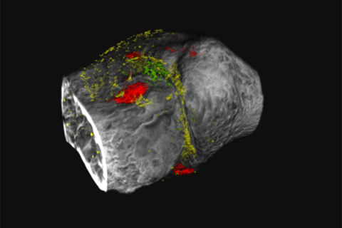
Medical imaging is no longer in Kansas, Toto, as a team led by Penn State researchers brings traditional black and white diagnostic images of X-rays and traditional CT scans into technicolor. The researchers developed novel contrast agents that target two proteins implicated in osteoarthritis, a degenerative joint disease commonly characterized as wear-and-tear arthritis. By marking the proteins with the contrast agents comprising newly designed metal nanoprobes, the researchers can use advanced imaging called “K-edge” imaging or photon-counting computed tomography (CT) to simultaneously track separate biological processes in color that, together, reveal more about the disease’s progression than a traditional scan.
Subtitle
Photon-counting CT scanner uses novel contrast agents in rats to observe multiple biological processes, revealing evidence of osteoarthritis long before clinical symptoms develop
Read the Full News Story

